Changing stroke rehab and research worldwide now.Time is Brain! trillions and trillions of neurons that DIE each day because there are NO effective hyperacute therapies besides tPA(only 12% effective). I have 523 posts on hyperacute therapy, enough for researchers to spend decades proving them out. These are my personal ideas and blog on stroke rehabilitation and stroke research. Do not attempt any of these without checking with your medical provider. Unless you join me in agitating, when you need these therapies they won't be there.

What this blog is for:

My blog is not to help survivors recover, it is to have the 10 million yearly stroke survivors light fires underneath their doctors, stroke hospitals and stroke researchers to get stroke solved. 100% recovery. The stroke medical world is completely failing at that goal, they don't even have it as a goal. Shortly after getting out of the hospital and getting NO information on the process or protocols of stroke rehabilitation and recovery I started searching on the internet and found that no other survivor received useful information. This is an attempt to cover all stroke rehabilitation information that should be readily available to survivors so they can talk with informed knowledge to their medical staff. It lays out what needs to be done to get stroke survivors closer to 100% recovery. It's quite disgusting that this information is not available from every stroke association and doctors group.

Wednesday, April 17, 2024

Informed consent requires clear communication in acute ischemic stroke

 With an objective damage diagnosis and EXACT REHAB PROTOCOLS THAT GET YOU TO 100% RECOVERY informed consent is a no-brainer, so solve this problem the correct way; 

  1. Objective damage diagnosis

  2. Exact 100% recovery protocols

Informed consent requires clear communication in acute ischemic stroke

Key takeaways:

  • Researchers conducted a literature review for informed consent with acute ischemic stroke.
  • Decisions should balance a patient’s wishes, decision of a surrogate and physicians’ knowledge of treatment options.

DENVER — In acute ischemic stroke requiring thrombolysis, informed consent requires communication and balance between the wishes of the patient, surrogates and physicians, according to a literature review.

“Any physician that is treating acute stroke patients at the bedside knows that there are perfect candidates for thrombolysis and less ideal candidates,” Alexis N. Kaiser, MD, resident appointee in the department of neurology at Indiana University School of Medicine, told Healio in a poster presentation at the American Academy of Neurology annual meeting.

Diverse group of health care professionals talking
Results of a literature review found that informed consent in acute ischemic stroke requires the patient, surrogates and physicians to communicate as much as possible to provide the clearest picture for treatment. Image: Adobe Stock

“We are put under time pressure to make these determinations very quickly.”

The American Academy of Neurology’s 2022 position statement on consent in case of acute stroke stipulated that verbal consent of patients “should be obtained and documented in the medical record by the treating physician.”

Kaiser and colleagues sought to review existing literature that related to ethical issues of informed consent in this patient population. Prior research shows a division among health care providers on the necessity of consent prior to thrombolysis, with 38% disagreeing that consent is necessary, while 33% of neurology trainees and 215 of neuro-related staff reported rigorous pursuit of informed consent prior to the procedure.

Their review encompassed 15 academic works that addressed the nature of consent when thrombolysis may be necessary with acute ischemic stroke, yielding three separate modes of thought:

  • Emergency consent, where the decision is made by a physician team when either the patient or surrogate is unable to participate;
  • Shared decision-making, which embraces a collaborative approach that matches best with the treatment plan as well as the patient’s wishes;
  • Informed consent, meaning the decision is made by the patient after receiving the best available evidence for intervention by the treating physician.

Additionally, Kaiser and colleagues’ review revealed perspectives from patients, surrogates and physicians, with opinions ranging from the patient’s view that acute stroke treatment is akin to more intensive responses like cardiopulmonary resuscitation and some patients might prefer death to disability; surrogates face uncertainty and a lack of preparation to make such a decision and advanced care documents lack nuance to properly guide their course of action; and care teams proceed with the mantra “time is brain” while attempting to balance mental and physical capacity assessments and legal ramifications.

The review further elucidated issues of framing discussions of care based on benefit/risk analyses, the lack of a standardized approach to treatment plans as well as perceptions of disability that differ between patient and physician.

“There is some discord in the field on ‘should we frame something positively or negatively, is that unethical?’” Kaiser noted. “It is our responsibility to give our best medical recommendation ... as long as the patient is able to ask questions.”

Reference:

Sattin JA, et al. Neurology. 2022:doi: 10.1212/WNL.000000000013040. Published Jan. 10, 2022. Accessed April 16, 2024.

Sources/Disclosures

Collapse

Source:

Kaiser AN, et al. Defining informed consent in acute ischemic stroke: Patient, surrogate decision maker and physician perspectives. Presented at: American Academy of Neurology annual meeting; April 13-18, 2024; Denver.
Disclosures: Kaiser reports no relevant financial disclosures.

International award for Box Hill stroke team


Anytime I see 'care' in any stroke press release I know the stroke medical world is not willing to disclose actual results because they are so fucking bad, it wouldn't look good, so misdirection is used. Don't fall for that misdirection! By touting 'care' they are not telling you about results or recovery which survivors want! Survivors don't care about door to needle time; you FUCKING BLITHERING IDIOTS; they want 100% recovery! why aren't you providing that?

Big fucking whoopee.


 But you tell us NOTHING ABOUT RESULTS. They remind us they 'care' about us multiple times but never tell us how many 100% recovered.  You have to ask yourself why they are hiding their incompetency by not disclosing recovery results.  ARE THEY THAT FUCKING BAD?


Three measurements will tell me if the stroke medical world is possibly not completely incompetent; DO YOU MEASURE ANYTHING?  I would start cleaning the hospitals by firing the board of directors, you can't let incompetency continue for years at a time.

There is no quality here if you don't measure the right things.

  1. tPA full recovery? Better than 12%?
  2. 30 day deaths? Better than competitors?
  3. rehab full recovery? Better than 10%?


You'll want to know results so call that hospital president(whomever that is) RESULTS are; tPA efficacy, 30 day deaths, 100% recovery. Because there is no point in going to that hospital if they are not willing to publish results.

In my opinion this partnership allows stroke hospitals to continue with their tyranny of low expectations and justify their complete failure to get survivors 100% recovered. Prove me wrong, I dare you in my stroke addled mind. If your stroke hospital goal is not 100% recovery you don't have a functioning stroke hospital.

All you ever get from hospitals are that they are following guidelines; these are way too static to be of any use. With thousands of pieces of stroke research yearly it would take a Ph.D. level research analyst to keep up, create protocols, and train the doctors and therapists in their use. 

If your stroke hospital doesn't have that, you don't have a well functioning stroke hospital, you have a dinosaur. 

Read up on the 'care' guidelines yourself. Survivors want RECOVERY not 'care'

“What's measured, improves.” So said management legend and author Peter F. Drucker 

The latest invalid chest thumping here:

International award for Box Hill stroke team

NTERVIEW OPPORTUNITY: Dr Philip Choi is available for interview upon request. Contact Marianna Alepidis to arrange a time

PHOTO: Available for download here
VIDEO FOOTAGE: Available for download here

  • The Eastern Health Box Hill Stroke Unit is being internationally recognised for meeting the highest standards in stroke treatment and care.
  • The award recognises both door to needle time and door in, door out time.
  • This year alone, Eastern Health Box Hill has treated 209 stroke patients.


The Eastern Health Box Hill Stroke Unit is being internationally recognised for meeting the highest standards in stroke treatment and care.

The World Stroke Organization (WSO) Angels has awarded the hospital WSO Gold Status.

Eastern Health Stroke Neurologist, Philip Choi said the award is an honour for the entire team who are known for having the nation’s fastest door-to-needle time.

“We are delighted to achieve WSO Gold Status and be recognised for our ongoing efforts to provide the community with the best stroke care possible,” Dr Choi said.

“This award acknowledges our commitment to excellence and the can-do attitude from everyone in the Stroke Team.”

Eastern Health Acute Stroke Nurse, Tanya Frost highlighted the performance of the team at the Box Hill Campus.

“Our greatest strength is our front end; our door to needle time, which is the time that the patient presents until the time they get thrombolysis and our door in, door out time. We're national leaders in both those times.

“We're very proud to say that we're the fastest in the country and are able to set that standard to ensure that patients receive excellent, prompt expert care, to aid the best possible recovery post stroke.”.

“We know that what we do today makes a massive difference to the patient and how they recover from their stroke,” Ms Frost explained.

The performance of the Hospital’s stroke unit was assessed as part of the Angels Initiative’s awards. The awards are given according to a set of stroke care key performance indicators. For WSO Gold status, this includes a target of restoring blood-flow to the brain to more than 50 per cent of eligible patients within 60 minutes of their hospital arrival.

This year alone, Eastern Health Box Hill has treated 209 stroke patients.

To achieve WSO Gold status, a hospital must demonstrate a range of outcomes, including optimum time to treatment, coordinated care, appropriate scans and screening, and ensuring patients are discharged from hospital on medications to minimise the risk of further stroke.

The Angels initiative, a partnership between the World Stroke Organization, European Stroke Organisation and Boehringer Ingelheim.


Contact details:

Marianna Alepidis

Media Officer

marianthe.alepidis@easternhealth.org.au

0437 875 825

Validity of an android device for assessing mobility in people with chronic stroke and hemiparesis: a cross-sectional study

 Assessments are completely worthless unless they point directly to the 100% recovery protocols. I see nothing here that suggests you go from the assessment to the chosen 100% recovery protocol. When the hell will the stroke medical world do ANYTHING TO GET STROKE SOLVED? I'd have you all fired! A lot of dead wood needs to removed in stroke and until that occurs stroke will never be solved!

Validity of an android device for assessing mobility in people with chronic stroke and hemiparesis: a cross-sectional study

Abstract

Background

Incorporating instrument measurements into clinical assessments can improve the accuracy of results when assessing mobility related to activities of daily living. This can assist clinicians in making evidence-based decisions. In this context, kinematic measures are considered essential for the assessment of sensorimotor recovery after stroke. The aim of this study was to assess the validity of using an Android device to evaluate kinematic data during the performance of a standardized mobility test in people with chronic stroke and hemiparesis.

Methods

This is a cross-sectional study including 36 individuals with chronic stroke and hemiparesis and 33 age-matched healthy subjects. A simple smartphone attached to the lumbar spine with an elastic band was used to measure participants’ kinematics during a standardized mobility test by using the inertial sensor embedded in it. This test includes postural control, walking, turning and sitting down, and standing up. Differences between stroke and non-stroke participants in the kinematic parameters obtained after data sensor processing were studied, as well as in the total execution and reaction times. Also, the relationship between the kinematic parameters and the community ambulation ability, degree of disability and functional mobility of individuals with stroke was studied.

Results

Compared to controls, participants with chronic stroke showed a larger medial-lateral displacement (p = 0.022) in bipedal stance, a higher medial-lateral range (p < 0.001) and a lower cranio-caudal range (p = 0.024) when walking, and lower turn-to-sit power (p = 0.001), turn-to-sit jerk (p = 0.026) and sit-to-stand jerk (p = 0.001) when assessing turn-to-sit-to-stand. Medial-lateral range and total execution time significantly correlated with all the clinical tests (p < 0.005), and resulted significantly different between independent and limited community ambulation patients (p = 0.042 and p = 0.006, respectively) as well as stroke participants with significant disability or slight/moderate disability (p = 0.024 and p = 0.041, respectively).

Conclusion

This study reports a valid, single, quick and easy-to-use test for assessing kinematic parameters in chronic stroke survivors by using a standardized mobility test with a smartphone. This measurement could provide valid clinical information on reaction time and kinematic parameters of postural control and gait, which can help(NOT GOOD ENOUGH! Come back when you have exact protocols for recovery!) in planning better intervention approaches. 

Introduction

Stroke is the second-leading cause of death in adults and represents a major cause of disability worldwide [1]. There are 12.2 million new cases of stroke each year, and 101 million people are living with the consequences of a stroke [1]. This number has almost doubled in the last 30 years [1]. It is noteworthy that 70% of strokes and 87% of stroke-related deaths and disability-adjusted life-years occur in low- and middle-income countries [2]. The impact of persistent stroke-related disability is significant to both individuals and society. Therefore, there is an ongoing need to address the challenges faced by stroke survivors [3]. Stroke typically causes sensorimotor deficits such as muscle weakness in the paretic limbs, impaired proprioceptive capabilities, sensory loss, vision problems, and spasticity [4]. These deficits affect the postural stability of stroke survivors and have significant influence on their mobility [5], causing functional mobility impairment after stroke [6].

Since functionality is related to daily life activities (i.e., sitting and standing, walking, turning, and going up and down stairs) [6], its impairment negatively affects the quality of life of post-stroke subjects [7]. Furthermore, functional mobility in the community requires not only the ability to maintain balance while walking and turning, but also to ambulate at 0.80 m/s or faster [8, 9]. Thus, difficulties with mobility control, particularly in balance and gait, are a priority clinical concern in this population [10].

In that sense, monitoring the improvement of functional mobility post-stroke is relevant as it could help to determine the effect of rehabilitation and thus expand therapeutic options [11,12,13]. The gold standard measurement of functional mobility is photogrammetry [14]. However, it is time-consuming and difficult to perform in clinical practice. Therefore, several tests have been used in clinical settings and research to determine functional mobility in stroke. Particularly, the Time Up and Go Test (TUG) [7], the Tinetti’s Scale of Mobility and Balance [7], the Dynamic Gait Index [15] and the Rivermead Mobility Index [16, 17]. These tools are quick and easy to use and do not require expensive materials. However, they provide a subjective evaluation and are less sensitive in detecting changes [18]. Therefore, incorporating instrumental systems into clinical assessments could improve the accuracy of results. These devices provide objective information on biomechanical aspects such as posture, movement, and compensatory strategies [18].

In this regard, Lin et al. [5] compared the results of clinical assessment scales (lower extremity subscale of the Fugl-Meyer Assessment, Berg Balance Scale and TUG) versus instrumental balance assessment (stability, proprioception and limits of stability) and showed that the later was superior to clinical alternatives in detecting balance impairments in stroke patients with mild balance disorders. As a conclusion, these authors recommended that clinicians consider the use of both classic clinical tests and quantitative biomechanical tools when evaluating stroke patients to improve the accuracy of assessments, leading to a better individualized treatment plan [5]. Similarly, the Stroke Recovery and Rehabilitation Roundtable of the International Stroke Recovery and Rehabilitation Alliance considered kinematic measures to be essential in assessing sensorimotor recovery [19]. Therefore, the use of a device that can help measure kinematic data when performing several daily activity tasks (such as rising, walking, turning, and sitting down) in a single test could help clinicians in evidence-based decision making. For such a purpose, current evidence examines various technological approaches. Many of them are not portable or easy to move, and the evaluation procedure is complex and time-consuming [20]. As a result, researchers have begun using smartphone applications to assess walking activity and balance performance in stroke [21].

Smartphones are equipped with advanced computing capabilities, global positioning system receivers, and sensing capabilities such as an inertial measurement unit (IMU), magnetometer, and barometer [22]. These features can also be found in wearable ambulatory monitors [22]. Hence, smartphones are currently gaining interest in post-stroke clinical assessment due to their validity, reliability, portability and price [21, 23], not only for measuring upper limb range of motion [24] but particularly for gait and balance assessment [21]. A systematic review of smartphone technology [25] found that a few systems have utilized IMU sensors embedded in smartphones, while more systems have integrated external sensors for data acquisition and used the smartphone as a data processing unit in stroke patients. For gait and balance assessment, smartphones has primarily been used to determine step counts and cadence [26, 27], recognize human movement activity [28], study posturography [20, 29], and analyze kinematic data during gait [23, 30, 31]. It should be noted that the current scientific evidence indicates that more studies are needed [20, 21], especially with standardized protocols and appropriate sample calculations [21]. To the best of our knowledge, no study has been found that uses a smartphone in a single test to evaluate kinematic data during the joint performance of different tasks relevant to activities of daily living, such as reaction time, sitting down, getting up or turning around, in stroke population. Therefore, this study aimed to assess the validity of using an Android device to evaluate kinematic data during the execution of a standardized mobility test that includes various tasks relevant to activities of daily living in people with chronic stroke and hemiparesis. To achieve this objective, the data registered by the sensor were compared between stroke and non-stroke individuals. In addition, the relationship between clinical tests and kinematic parameters were studied for patients. If our working hypothesis is met, the inclusion of an inertial sensor on an Android device when performing a standardized mobility test will provide further information about which daily activity tasks have greater impairment in the population with chronic stroke, without the need to allocate more time for the clinical assessment.

More at link.

HIV Drugs May Reduce Alzheimer’s Risk

 FYI.

Your risk of dementia, has your doctor told you of this?  Your doctor is responsible for preventing this!

1. A documented 33% dementia chance post-stroke from an Australian study?   May 2012.

2. Then this study came out and seems to have a range from 17-66%. December 2013.`    

3. A 20% chance in this research.   July 2013.

4. Dementia Risk Doubled in Patients Following Stroke September 2018 

The latest here:


HIV Drugs May Reduce Alzheimer’s Risk

Summary: Common HIV drugs could reduce the incidence of Alzheimer’s disease (AD). Utilizing anonymized prescription data from over 225,000 individuals, the study found that HIV-positive patients taking reverse transcriptase (RT) inhibitors showed a significantly lower rate of AD compared to the general population.

This discovery builds on previous findings that Alzheimer’s-linked genes might be recombined by enzymes similar to those targeted by HIV treatments. The results could pave the way for new therapeutic strategies using existing drugs to combat the growing AD crisis.

Key Facts:

  1. The study analyzed prescription data from 225,000 individuals, revealing that HIV-positive patients over 60 taking RT inhibitors had fewer Alzheimer’s diagnoses compared to their non-HIV counterparts.
  2. RT inhibitors, initially developed for HIV, might inhibit similar enzymes in the brain, suggesting a potential mechanism for their effect on Alzheimer’s disease.
  3. The research was supported by notable foundations and the NIH, highlighting its credibility and the significant interest in translating these findings into new treatments for AD.

Source: Sanford Burnham Prebys

Alzheimer’s disease (AD) currently afflicts nearly seven million people in the U.S. With this number expected to grow to nearly 13 million by 2050, the lack of meaningful therapies represents a major unmet medical need. Scientists at Sanford Burnham Prebys have now identified promising real-world links between common HIV drugs and a reduced incidence of AD.

The study, led by Jerold Chun, M.D., Ph.D., was published in Pharmaceuticals.

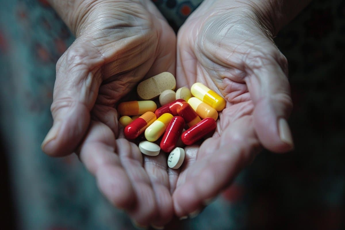
This shows a person holding pills.
The brain appears to have its own RTs that are different from those in viruses, and the research team wondered if inhibiting brain RTs with HIV drugs actually helps AD patients. Credit: Neuroscience News

Chun’s new research builds on his lab’s landmark publication in Nature in 2018 that described how somatic gene recombination in neurons can produce thousands of new gene variants within Alzheimer’s disease brains. Importantly, it also revealed for the first time how the Alzheimer’s-linked gene, APP, is recombined by using the same type of enzyme found in HIV. 

The enzyme, called reverse transcriptase (RT), copies RNA molecules and changes them into complementary DNA duplicates that can then be inserted back into DNA, producing permanent sequence changes within the cell’s DNA blueprint.

HIV and many other viruses rely on RT to hijack a host’s cells to establish a chronic infection, so drugs that block the RT enzyme’s activity have become a common part of treatment cocktails for keeping HIV at bay.

The brain appears to have its own RTs that are different from those in viruses, and the research team wondered if inhibiting brain RTs with HIV drugs actually helps AD patients.

To assess the link between real-world RT inhibitor exposure and AD in humans, the team analyzed anonymized medical records with prescription claims from more than 225,000 control and HIV-positive patients, and found that RT inhibitor exposure was associated with a statistically significant reduced incidence and prevalence of AD.

“Thus, we looked at HIV-positive individuals taking RT inhibitors and other combined antiretroviral therapies as they aged, and asked the question: How many of them got Alzheimer’s disease?” says Chun.

“And the answer is that there were many fewer than might have been expected compared to the general population.”

Of the more than 225,000 individuals with claims data in the study, just shy of 80,000 were HIV-positive individuals over the age of 60. More than 46,000 had taken RT inhibitors during a nearly three-year observation period from 2016 to 2019. The data was obtained through a collaboration with health information technology and clinical research firm IQVIA, led by Tiffany Chow, M.D.

In living persons with HIV, there were 2.46 Alzheimer’s disease diagnoses per 1,000 persons among HIV-positive individuals taking these inhibitors, versus 6.15 for the general population.

This control group was represented by more than 150,000 HIV-negative patients over the age of 60 with medical insurance claims related to treatment for the common cold.

“You cannot feasibly run a prospective clinical trial with this number of patients,” Chun adds. “This approach is a way to look at how a drug can act on a large patient population.”

Chun underscores that the drugs patients took in this retrospective study were designed to counter RT activity in HIV and likely only had a limited effect on many different possible forms of the enzyme active in the brain.

“What we’re looking at now is very crude,” says Chun. “The clear next step for our lab is to identify which versions of RTs are at work in the AD brain so that more targeted treatments can be discovered, while prospective clinical trials of currently available RT inhibitors on persons with early AD should be pursued.”

Jerold Chun, M.D. Ph.D., is a professor in the Center for Genetic Disorders and Aging Research at Sanford Burnham Prebys.

Additional authors on the study include Tiffany W. Chow, Mark Raupp, Matthew W. Reynolds, Siying Li and Gwendolyn E. Kaeser.

Funding: The work was supported by the National Institute on Aging – NIH (R01AG071465, R01AG065541 and R56AG073965), the Shaffer Family Foundation and the Bruce Ford & Anne Smith Bundy Foundation.

About this Alzheimer’s disease and neuropharmacology research news

Author: Greg Calhoun
Source: Sanford Burnham Prebys
Contact: Greg Calhoun – Sanford Burnham Prebys
Image: The image is credited to Neuroscience News

Use of functional magnetic resonance imaging to identify cortical loci for lower limb movements and their efficacy for individuals after stroke

 Ask your competent? doctor EXACTLY HOW THIS WILL GET YOU RECOVERED!  Because I see nothing useful.

Use of functional magnetic resonance imaging to identify cortical loci for lower limb movements and their efficacy for individuals after stroke

Abstract

Background

Identification of cortical loci for lower limb movements for stroke rehabilitation is crucial for better rehabilitation outcomes via noninvasive brain stimulation by targeting the fine-grained cortical loci of the movements. However, identification of the cortical loci for lower limb movements using functional MRI (fMRI) is challenging due to head motion and difficulty in isolating different types of movement. Therefore, we developed a custom-made MR-compatible footplate and leg cushion to identify the cortical loci for lower limb movements and conducted multivariate analysis on the fMRI data. We evaluated the validity of the identified loci using both fMRI and behavioral data, obtained from healthy participants as well as individuals after stroke.

Methods

We recruited 33 healthy participants who performed four different lower limb movements (ankle dorsiflexion, ankle rotation, knee extension, and toe flexion) using our custom-built equipment while fMRI data were acquired. A subgroup of these participants (Dataset 1; n = 21) was used to identify the cortical loci associated with each lower limb movement in the paracentral lobule (PCL) using multivoxel pattern analysis and representational similarity analysis. The identified cortical loci were then evaluated using the remaining healthy participants (Dataset 2; n = 11), for whom the laterality index (LI) was calculated for each lower limb movement using the cortical loci identified for the left and right lower limbs. In addition, we acquired a dataset from 15 individuals with chronic stroke for regression analysis using the LI and the Fugl–Meyer Assessment (FMA) scale.

Results

The cortical loci associated with the lower limb movements were hierarchically organized in the medial wall of the PCL following the cortical homunculus. The LI was clearer using the identified cortical loci than using the PCL. The healthy participants (mean ± standard deviation: 0.12 ± 0.30; range: – 0.63 to 0.91) exhibited a higher contralateral LI than the individuals after stroke (0.07 ± 0.47; – 0.83 to 0.97). The corresponding LI scores for individuals after stroke showed a significant positive correlation with the FMA scale for paretic side movement in ankle dorsiflexion (R2 = 0.33, p = 0.025) and toe flexion (R2 = 0.37, p = 0.016).

Conclusions

The cortical loci associated with lower limb movements in the PCL identified in healthy participants were validated using independent groups of healthy participants and individuals after stroke. Our findings suggest that these cortical loci may be beneficial(NOT GOOD ENOUGH! Come back when you have exact protocols for recovery!) for the neurorehabilitation of lower limb movement in individuals after stroke, such as in developing effective rehabilitation interventions guided by the LI scores obtained for neuronal activations calculated from the identified cortical loci across the paretic and non-paretic sides of the brain.

Background

Functional magnetic resonance imaging (fMRI) has been widely used to investigate the motor functions of the human brain, particularly upper limb movements such as finger tapping and hand grasping/clenching. Distinct cortical loci have been identified for different types of upper limb movement [1,2,3], and these cortical loci have been successfully employed for the neurorehabilitation/neuroplasticity of individuals in the chronic stage after stroke [4,5,6]. The importance of identifying the cortical loci for lower limb movement in stroke rehabilitation has been discussed in previous studies [7, 8]. For example, noninvasive brain stimulations such as repetitive transcranial magnetic stimulation (rTMS) and transcranial direct current stimulation (tDCS) have been demonstrated to be effective in improving the gait and balance performance of individuals with subacute and chronic stroke by targeting the cortical loci, including the primary motor cortex (M1) [7]. We believe rehabilitation outcomes would be further enhanced by targeting the stimulation of fine-grained cortical loci for lower limb movement.

In this context, previous studies have investigated neuronal patterns observed from fMRI during lower limb movements [9,10,11,12]. For example, Luft and colleagues (2002) compared brain activations between upper and lower limb movements by incorporating finger, elbow, and knee movements in their lateralization index (LI) across various regions-of-interest (ROIs), including the M1, primary somatosensory cortex (S1), primary motor area (SMA), and cerebellum [10]. Kapreli et al. extended these findings by including the ankle and toes to differentiate the LI of brain activations for finger movement from that for the movement of lower limb joints [11, 12] and by combining movements across the ankle, knee, and hip [9]. These previous studies have generally reported different activation loci for lower limb movements compared with upper limb movements and overlapping spatial layouts for neuronal activations across lower limb movements. Another line of research has compared the neuronal activations of imagined lower limb movements with executed and/or observed movements for the right ankle [13], foot-kicking [14], and stepping [15].

However, few studies have investigated the distinct cortical loci considering hierarchical representations in the cortical homunculus for the movement of the ankle, toe, or knee, which are feasible movements of the lower limb extremities for fMRI acquisition because head motion is potentially more controllable compared with hip joint movement [11]. The identification of cortical loci specific to these lower limb movements in the median wall of the sensorimotor area mainly in the paracentral lobule (PCL) region is more challenging than for upper limb movements because the motor cortex associated with the lower limbs is smaller in volume than that for the upper limbs based on the cortical homunculus [16]. In addition, isolation of individual lower limb movements is more demanding due to the potentially greater head motion [10, 17,18,19].

Previous studies have investigated the neuronal activation patterns of lower limb movements based on the guidance of MR-compatible equipment. These studies include the identification of neuronal activation patterns for active and passive stepping movements [15, 20, 21] and pedaling [18] and the real-time monitoring of ankle, knee, and hip torques with their associated neuronal activations [22]. In the present study, we developed a custom-made MR-compatible footplate and leg cushion to isolate individual lower limb movements and minimize potential head motion during fMRI data acquisition. We then identified the cortical loci for lower limb movements using fMRI data acquired from healthy participants and subsequent multivariate analysis. Conventionally, it is not easy to delineate these loci due to the constrained cortical regions, particularly in the PCL, which is the medial continuation of the precentral and postcentral gyri [23], and overlapping functional territories across the lower limbs. We also investigated the efficacy of the cortical loci identified from healthy participants for individuals with chronic stroke. We hypothesized that our custom-built equipment and multivariate analytical methods would be useful for identifying hierarchically organized cortical loci for lower limb movements (i.e., an inferior location for the toe to a superior location for the knee). We also hypothesized that the identified cortical loci would be useful in evaluating the neural features of individuals with chronic stroke.

More at link.

Tuesday, April 16, 2024

The effects of genetic and modifiable risk factors on brain regions vulnerable to ageing and disease

Well, I don't have diabetes or pre-diabetes and don't live anywhere close to air pollution. Alcohol is used to vastly increase my social participation which is going to prevent dementia, so there! Don't follow me.

The effects of genetic and modifiable risk factors on brain regions vulnerable to ageing and disease

Abstract

We have previously identified a network of higher-order brain regions particularly vulnerable to the ageing process, schizophrenia and Alzheimer’s disease. However, it remains unknown what the genetic influences on this fragile brain network are, and whether it can be altered by the most common modifiable risk factors for dementia. Here, in ~40,000 UK Biobank participants, we first show significant genome-wide associations between this brain network and seven genetic clusters implicated in cardiovascular deaths, schizophrenia, Alzheimer’s and Parkinson’s disease, and with the two antigens of the XG blood group located in the pseudoautosomal region of the sex chromosomes. We further reveal that the most deleterious modifiable risk factors for this vulnerable brain network are diabetes, nitrogen dioxide – a proxy for traffic-related air pollution – and alcohol intake frequency. The extent of these associations was uncovered by examining these modifiable risk factors in a single model to assess the unique contribution of each on the vulnerable brain network, above and beyond the dominating effects of age and sex. These results provide a comprehensive picture of the role played by genetic and modifiable risk factors on these fragile parts of the brain.